The Implantation of the AbioCor Artificial Heart
2001 · Louisville, United States
The world's first self-contained artificial heart, the AbioCor, was implanted in a patient at Jewish Hospital in Louisville, Kentucky.
July 19, 1983
The first three-dimensional reconstruction of a human head in a CT scan was published, marking a milestone in medical imaging technology.
San Francisco, United States | University of California, San Francisco
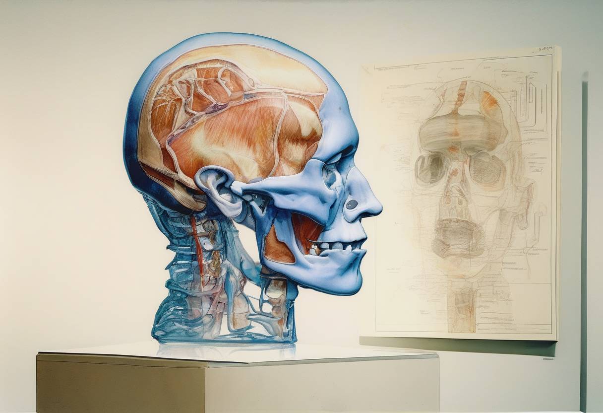
On July 19, 1983, a groundbreaking advancement in medical imaging technology was achieved with the publication of the first three-dimensional reconstruction of a human head using computed tomography (CT) scans. This milestone marked a significant leap forward in the field of diagnostic imaging and had profound implications for medicine and surgery.
Computed tomography, or CT scanning, was developed in the early 1970s and quickly became a vital tool in medical diagnostics. Traditional CT scans provided two-dimensional cross-sectional images of the body, which were invaluable for identifying and diagnosing various conditions. However, the ability to visualize structures in three dimensions was limited.
The 1983 breakthrough involved the use of advanced computer algorithms to reconstruct a series of two-dimensional CT images into a three-dimensional model. This process allowed for a more comprehensive visualization of the human anatomy, providing clinicians with a clearer understanding of complex structures and spatial relationships within the body.
The reconstruction was achieved by a team of researchers led by Dr. Michael Vannier, Dr. James Marsh, and Dr. John Warren at the Mallinckrodt Institute of Radiology at Washington University in St. Louis. Their work demonstrated the potential of 3D imaging to enhance diagnostic accuracy and improve surgical planning.
The introduction of 3D CT scan reconstructions revolutionized medical imaging by:
Improving Diagnostic Precision: Clinicians could now view anatomical structures in three dimensions, leading to more accurate diagnoses and better treatment planning.
Enhancing Surgical Planning: Surgeons gained the ability to visualize the operative field in 3D, allowing for more precise and less invasive procedures.
Advancing Research and Education: The technology provided a new tool for medical research and education, offering detailed anatomical models for study and training.
The success of the first 3D CT scan reconstruction paved the way for further advancements in imaging technology. It led to the development of more sophisticated imaging techniques, including magnetic resonance imaging (MRI) and positron emission tomography (PET) scans, which also adopted 3D reconstruction methods.
Today, 3D imaging is a standard practice in medical diagnostics and treatment, playing a crucial role in fields such as oncology, cardiology, and orthopedics. The pioneering work of Vannier and his colleagues continues to impact the medical field, underscoring the importance of innovation in improving patient care.
In summary, the publication of the first three-dimensional reconstruction of a human head using CT scans on July 19, 1983, was a landmark event in medical history, setting the stage for the modern era of medical imaging.
Source: en.wikipedia.org